Mail Us
contact@biedx.com
contact@biedx.com

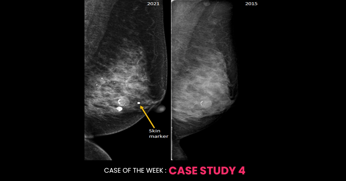
Underneath the area of the palpable lump in the left breast, a circumscribed isodense mass is seen with internal coarse calcifications; most likely an involuting fibroadenoma. It was also seen in the 2015 study with peripheral calcification. Though the patient may be now able to feel the same lesion (may be due to post-menopausal changes in the breast or maybe the patient has started consciously performing breast self-examination etc.) at the same time a new underlying more sinister cause that is obscured by dense breast tissue on mammogram is not ruled out, she deserves further evaluation with cone compression view/ Tomosynthesis and ultrasound for the region of new palpable concern. That is why this mammogram should be reported as BI-RADS 0 and the patient should be further evaluated. If the further workup confirms the palpable lump to correlate with this involuting fibroadenoma and no other suspicious feature is identified, then she can be discharged with final BI-RADS 2 after the complete work up.
It will be a good idea to read the “Appropriateness criteria for palpable breast masses: Slanetz PJ, Moy L, Baron P, Didwania AD, Heller SL, Holbrook AI, Lewin AA, Lourenco AP, Mehta TS, Niell BL, Stuckey AR. ACR appropriateness criteria® breast imaging of pregnant and lactating women. Journal of the American College of Radiology. 2018 Nov 1;15(11):S263-75.
If imaging studies raise suspicion, a biopsy may be performed to obtain a tissue sample for histopathological examination. Types of biopsies include:
If imaging studies raise suspicion, a biopsy may be performed to obtain a tissue sample for histopathological examination. Types of biopsies include:
Regular breast self-examinations and screening mammograms are crucial for early detection of breast abnormalities. Women are encouraged to become familiar with the normal look and feel of their breasts to identify any changes early and seek medical evaluation promptly.
In conclusion, while palpable breast masses can be alarming, they are often benign. However, due to the potential risk of malignancy, it is essential to evaluate any new or changing breast masses thoroughly through clinical examination, imaging, and biopsy when necessary.
To provide state-of-the-art education and career growth to radiologists and surgeons for improved patient care through wide availability of highly skilled doctors.
Copyright © 2025 All rights reserved.